


Page 6 - Ärzteblatt Mecklenburg-Vorpommern, Oktober 2026
P. 6
WISSENSCHAFT UND FORSCHUNG


           den letzten Jahrzehnten kein wesentlicher Rückgang der In-  FAVORS – zentrale Projektidee
           suffizienzraten zu verzeichnen. Dies verdeutlicht, dass techni-
           sche Optimierungen der verfügbaren Verfahren allein nicht   Die grundlegende Neuerung des Projektes FAVORS besteht in
           ausreichen und ein erheblicher Bedarf an verbesserten Stra-  einem Paradigmenwechsel der Überwachung. Bisherige An-
           tegien zur frühzeitigen Diagnostik besteht. Ein solches Verfah-  sätze zur Erkennung einer Anastomoseninsuffizienz messen
           ren zur direkten, kontinuierlichen Diagnostik von Anastomo-  fast immer indirekt über systemische Entzündungsmarker
           senkomplikationen stehen bislang nicht zur Verfügung und   oder  über  Drainageflüssigkeit.  Sie  sprechen  damit  erst  an,
           waren der ausschlaggebende Punkt hinter der Idee dieses   wenn eine systemische Reaktion auftrat oder eine definitive
           Forschungsprojektes.                                 Dehiszenz vorliegt. FAVORS jedoch verlagert die Messung


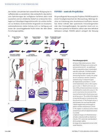
                                                                                      Forschungsprojekt:
                                                                                      A) Sensor fabrication process. Silver
                                                                                      and PEDOT:PSS layers are screen-prin-
                                                                                      ted and dried at 50°C. An additional
                                                                                      PDO encapsulation layer is laminated
                                                                                      onto the substrate. Finished samples
                                                                                      are cut using a high-precision laser
                                                                                      structuring machine. Finally, the samp-
                                                                                      les are contacted, and the contact is
                                                                                      isolated using 2-component glue.
                                                                                      B) Implantation procedure: 1: Envisi-
                                                                                      oned implantation sites at the esopha-
                                                                                      gus or colon. 2: Sketch and cross-sec-
                                                                                      tion of the printed anastomosis sensors,
                                                                                      red dashed line marks the cutout 3: The
                                                                                      anvil is inserted at the oral end of the
                                                                                      resected bowel and is fixed using a pur-
                                                                                      se-string suture. The shaft of the stapler
                                                                                      is inserted through an enterotomy into
                                                                                      the aboral end. 4: The finished sensor is
                                                                                      slid between the anvil and the shaft.
                                                                                      5: Both stapler ends are brought to-
                                                                                      gether and the anastomosis is fired.
                                                                                      6: The stapler is removed, and an intact
                                                                                      anastomosis with an implanted sensor
                                                                                      is created.
                                                                                      C) Optical images of a finished sensor
                                                                                      already contacted with an Amphenol
                                                                                      Clincher FPC connector and standard
                                                                                      20 cm jumper cables.
                                                                                      D) Image of an implanted sensor in the
                                                                                      small intestine.


       Seite 358                                                                     ÄRZTEBLATT MECKLENBURG-VORPOMMERN

